Xenium® Spatial Analyzer | BioChain Institute, Inc.
Xenium® Prime is precision insights.
Xenium Prime 5K assay from 10x Genomics is specifically designed for single-cell resolution capturing of a much larger number of genes than traditional technologies. This makes it ideal for more in-depth cellular analysis exploring heterogeneity of gene expression across tissue types like tumors, immune landscapes, or in neuroscientific studies. It offers single-cell spatial imaging of up to 5,000 genes across entire tissue sections.
This platform utilizes the Xenium Prime 5K pan-tissue and pathways assays, which are expertly curated panels enabling comprehensive exploration of biological materials with exceptional sensitivity, specificity, and spatial fidelity. Customizable for up to 50 to 100 additional genes, Prime allows tailored analyses to meet specific research needs.
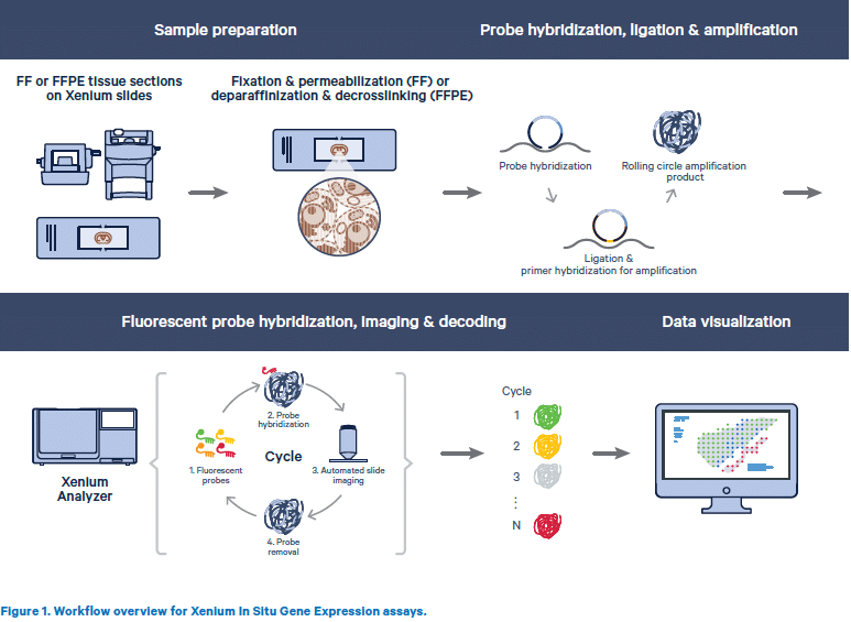
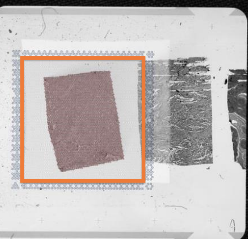
– Tissue samples are sectioned onto a Xenium slide to begin the workflow.
– Sections are treated to preserve RNA and allow access for circularizable DNA probes.
– Each DNA probe consists of two independent regions that hybridize to the target RNA and contain a gene-specific barcode sequence.
– The probe ends are ligated to form a circular DNA probe, which is then enzymatically amplified for detection.
– If one part of the probe binds off-target, ligation does not occur, preventing false signals and ensuring high specificity.
– The platform will later support DNA-barcoded antibodies for multiplexed protein detection in tissue sections.
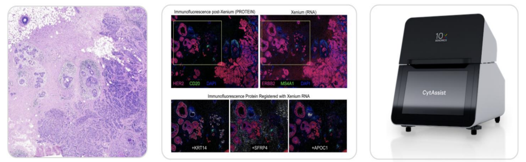
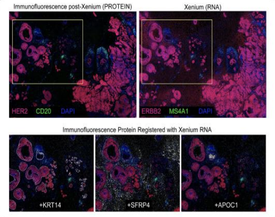
– Insights are maximized using post Xenium processing such as H&E or IF staining and Visium on the same section.
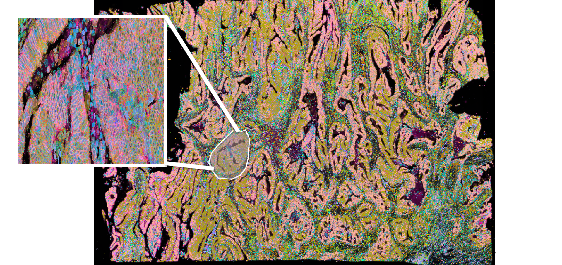
– Xenium transcriptional data can be directly compared to morphological data from H&E and IF staining.