Visium® Spatial Gene Expression | BioChain Institute, Inc.
Visium HD is unbiased discovery.
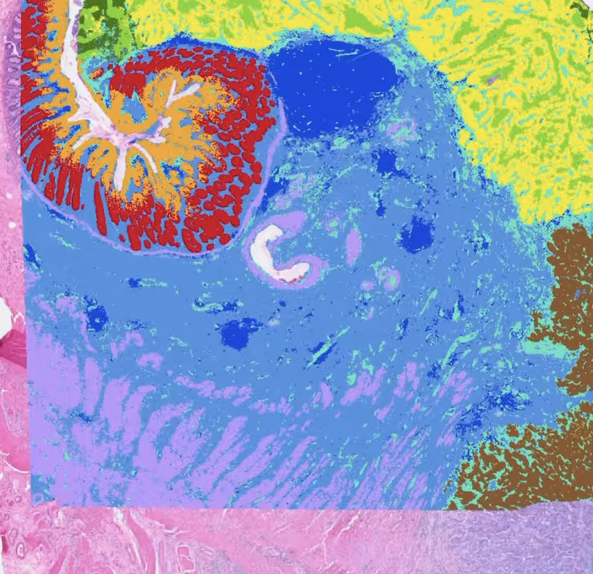
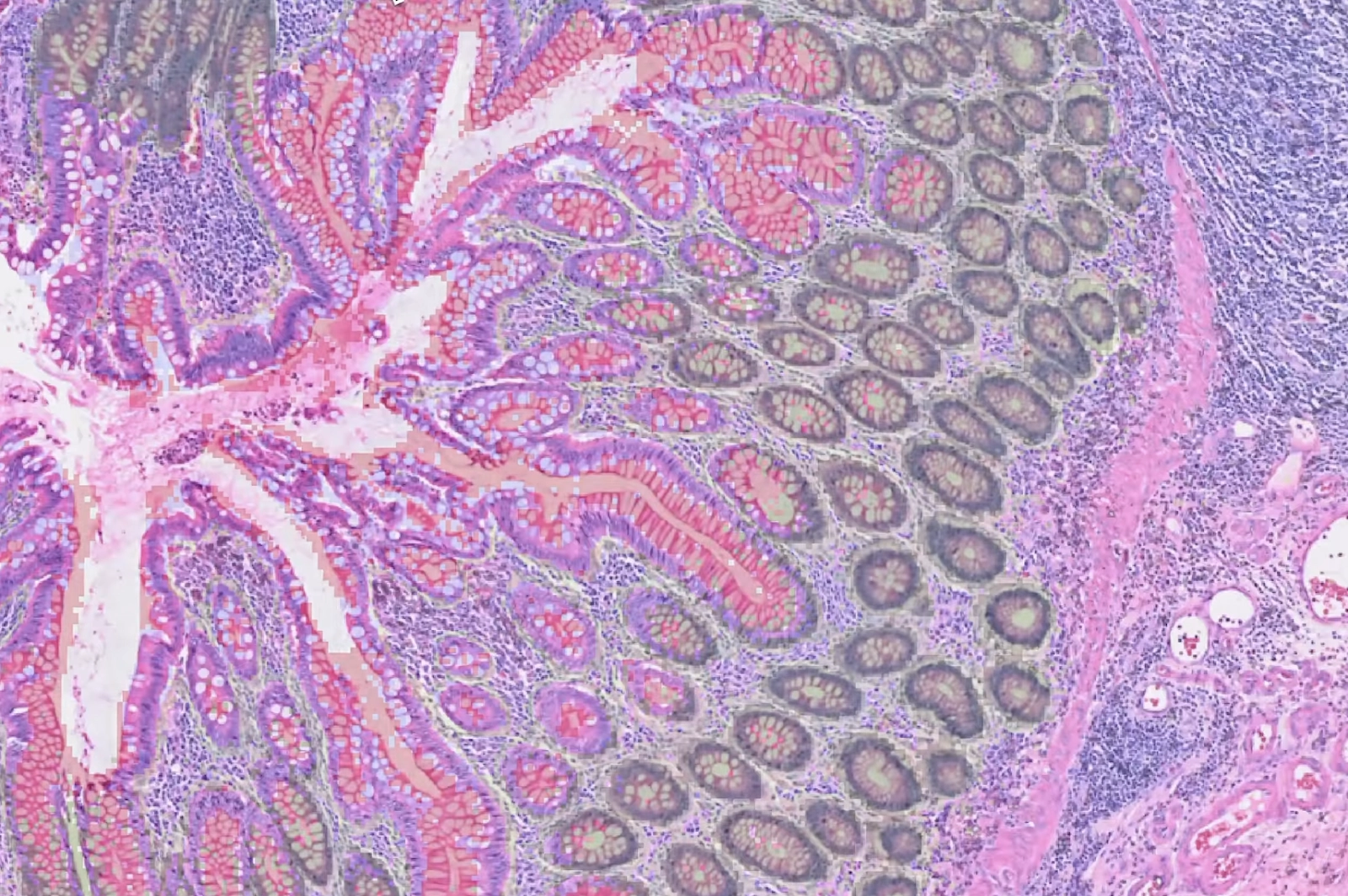
Building upon the original Visium® platform, Visium® HD features a continuous lawn of oligonucleotides arrayed in millions of barcoded squares without gaps, achieving single cell–scale spatial resolution. This design allows for precise visualization of whole transcriptome and tissue morphology with uninterrupted coverage. With exceptional sensitivity and the integration of the Visium CytAssist technology, Visium® HD enables detection of low-abundance genes and facilitates seamless transfer of gene expression probes from tissue sections onto Visium HD slides, ensuring accurate spatial data capture.
Building on the 10x Visium Digital Spatial Gene Expression kit, Visium HD has exciting new features that open spatial assay horizons. Map the whole transcriptome within the tissue context at high-definition 2um resolution. Unravel biological architectures in normal and diseased tissue and discover new biomarkers. Visualize the spatial organization of awe-inspiring cell types, states, and biomarkers
Our ready-to-use, robust workflow smoothly integrates into current laboratory methods and tools for whole tissue section analysis.
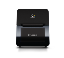
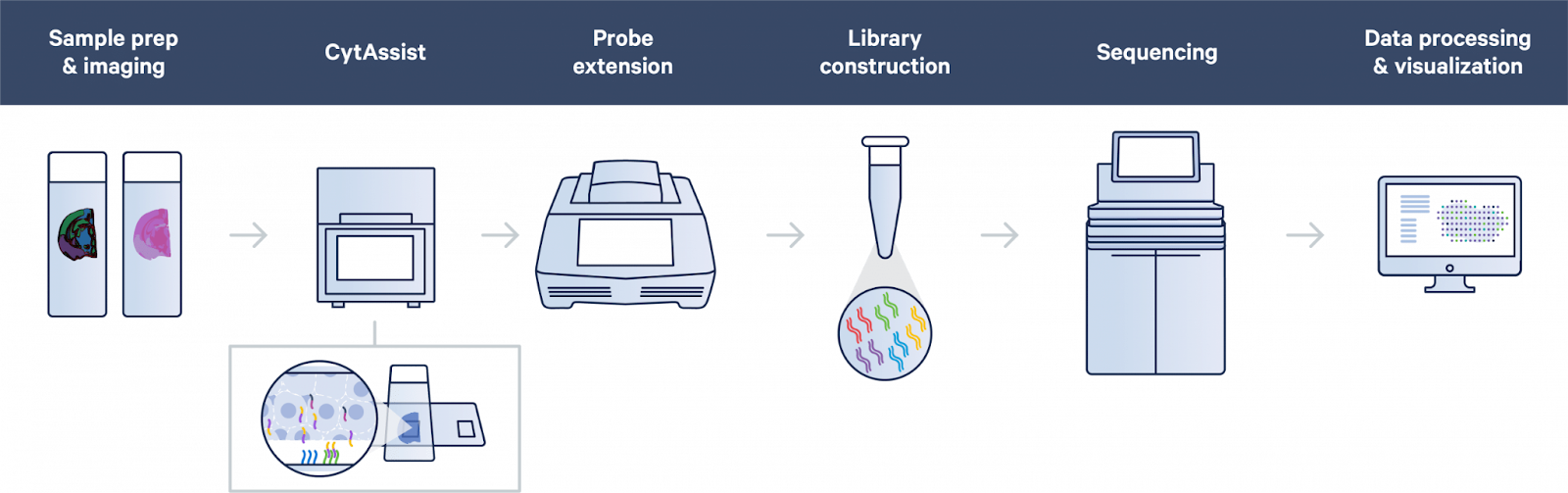
Facilitate transfer of transcriptomic probes in FFPE or fresh frozen samples with Visium CytAssist. In the Visium CytAssist workflow, sectioning, tissue preparation, staining (H&E or IF), and imaging take place on a standard glass slide.
After probe hybridization, two standard glass slides and a Visium slide with two Capture Areas are placed in the CytAssist instrument. A brightfield image is captured to provide spatial orientation for data analysis.
The tissue sections on the standard slides are aligned on top of the Capture Areas, where transcriptomic probes hybridize to the Visium slide.
Once hybridized, the remaining steps—starting with probe extension—follow the standard Visium workflow outside the CytAssist instrument.
The tissue sections are permeabilized, releasing ligated probe pairs that bind to adjacent capture probes on the slide, enabling gene expression capture.
Gene-specific probe pairs hybridize to their target transcript and are then ligated together.
The ligated probes are extended to incorporate spatial barcodes, and sequencing libraries are prepared.
Fresh-Frozen Tissue Workflow
In fresh-frozen tissues, RNA is released by fixation and permeabilization, binding to adjacent capture probes for gene expression capture.
Sequencing libraries are synthesized and captured, enabling spatial transcriptomic analysis.
With 10x Visium Digital Spatial Gene Expression, you can gain a holistic view of disease complexity, discover new biomarkers, map the spatial organization of cell atlases, and identify spatiotemporal gene expression patterns.
Reach out about your project with one click! Address all of your concerns or questions with our live spatial consulting to see what is best for you.